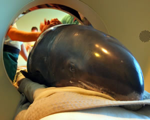
A CT scan was performed on the pygmy killer whale 'Pete'. credit Marc M. Ellis/Mote Marine Laboratory
The condition of the pygmy killer whales brought to Mote's Dolphin and Whale Hospital on June 16 after stranding near Boca Grande continues to improve, though both are still considered to be in critical condition. One of the whales, nicknamed "Pete" after the boat captain who found him, underwent a CT scan Thursday evening at Axcess Diagnostics in Sarasota.
The CT scan – read by neuroradiologist Dr. Paul Macchi of Axcess Diagnostics ‑ showed no abnormalities in the animal's brain, but it did indicate there were lesions on the animal's lungs, said Mote veterinarian Dr. Charles Manire. "The CT scan helped confirm what we believed: that there were lesions – possibly from parasites ‑ in the animal's lungs," Manire said. "This information will help us provide a better treatment plan for the animals."
A CT, or computer tomography, scan combines a sophisticated X-ray system with a high-speed computer to give a diagnosing physician an unobstructed look at organs and other structures that cannot be seen in traditional X-rays. Thursday's CT scan on Pete followed a June 18 MRI on Dallas.
Manire chose to do a CT scan this time because he thought it would offer better results in this situation. "An MRI provides the best images of the brain," Manire said. "But you have to be very still. Dolphins and small whales breathe two or three times per minute and they move their heads when they do so, so it's very difficult to keep them still. A CT scan, on the other hand, takes only a few seconds to complete, so movement isn't as much of an issue. I just can't thank Dr. Miley and his staff enough for providing this tool and their expertise to help us treat Dallas and Pete."
Axcess Diagnostics, owned by Dr. Stephen Miley, CEO, donated staff time and the use of the CT equipment to Mote.